NEUROANATOMY
AN ATLAS OF STRUCTURES SECTIONS, AND SYSTEMS
ISBN: 9780781763288
Συγγραφέας: HAINES DUANE
Κωδικός: 9780781763288
Μη διαθέσιμο
Τιμή
42,00€ 15,00€
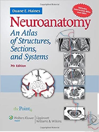
Now in its 25th year, this best-selling work is the only neuroanatomy atlas to integrate neuroanatomy and neurobiology with extensive clinical information. It combines full-color anatomical illustrations with over 200 MRI, CT, MRA, and MRV images to clearly demonstrate anatomical-clinical correlations. This edition contains many new MRI/CT images and is fully updated to conform to Terminologia Anatomica. Fifteen innovative new color illustrations correlate clinical images of lesions at strategic locations on pathways with corresponding deficits in Brown-Sequard syndrome, dystonia, Parkinson disease, and other conditions. The question-and-answer chapter contains over 235 review questions, many USMLE-style.
| Χαρακτηριστικά Προϊόντος | |
|---|---|
| ISBN | 9780781763288 |
| Συγγραφέας | HAINES DUANE |
| Εκδότης | LIPPINCOTT WILLIAMS AND WILKINS |
| Επίπεδο | ΠΑΝΕΠΙΣΤΗΜΙΟ |
| Εξώφυλλο | ΜΑΛΑΚΟ |
| Αρ. Έκδοσης | 7η |
| Έτος Έκδοσης | 2008 |
| Σελίδες | 341 |
| Χώρα προέλευσης | Η.Π.Α |
Αυτή η σελίδα προστατεύεται από το σύστημα reCAPTCHA της Google. Μάθετε περισσότερα.


