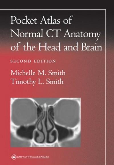
POCKET ATLAS OF NORMAL CT ANATOMY OF THE HEAD AND BRAIN
ISBN: 9780781729499
Συγγραφέας: MICHELLE M. SMITH
Κωδικός: 9780781729499
Άμεση παραλαβή / Παράδοση σε 1-3
Τιμή
20,00€ 10,00€
Featuring 73 sharp, new images obtained with state-of-the-art scanning technology, the Second Edition of this popular pocket atlas is a quick, handy guide to interpreting computed tomography images of the brain and calvarium, temporal bone, orbit, nasal cavity, and paranasal sinuses. The book helps readers recognize normal anatomic structures on CT scans and distinguish these structures from artifacts.Each page presents a high-resolution CT scan, with anatomic landmarks clearly labeled. Directly above the scan are a key to the labels and a thumbnail illustration that orients the reader to the plane of view (sagittal, axial, or coronal).
| Χαρακτηριστικά Προϊόντος | |
|---|---|
| ISBN | 9780781729499 |
| Συγγραφέας | MICHELLE M. SMITH |
| Εκδότης | LIPPINCOTT WILLIAMS AND WILKINS |
| Επίπεδο | ΠΑΝΕΠΙΣΤΗΜΙΟ |
| Εξώφυλλο | ΜΑΛΑΚΟ |
| Αρ. Έκδοσης | 2η |
| Έτος Έκδοσης | 2001 |
| Σελίδες | 79 |
| Χώρα προέλευσης | Η.Π.Α |
Αυτή η σελίδα προστατεύεται από το σύστημα reCAPTCHA της Google. Μάθετε περισσότερα.


